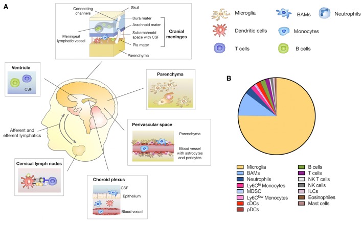
Figure 1.
Immune compartments in the CNS. (A) The brain parenchyma is tightly shielded from the systemic immune system by the BBB and BCB that restricts the entry of peripheral immune and inflammatory cells. Yolk sac-derived microglia represent in the innate immune cell type within the brain parenchyma and exert key functions in host defense, immune surveillance and tissue homeostasis. In addition to the brain parenchyma, the CNS comprises structural interfaces that connect the CNS with the periphery, such as the choroid plexus, the meninges and the perivascular spaces. Border-associated macrophages (BAMs) are located in each of these niches. Circulating immune cells such as dendritic cells (DC), B- and T-cells are found in the CSF predominately in the meninges, ventricles and choroid plexus. A meningeal lymphatic system allows DC trafficking from the CNS to CNS draining lymph nodes i.e., cervical lymph nodes. Direct channels that connect the skull bone marrow with the dura enable neutrophils to migrate to the CNS as immediate responders to CNS damage. (B) Relative amount of distinct CNS immune cell populations located in the parenchyma and border areas of the CNS. Depicted amounts are based on data published in Mrdjen et al. (20).

