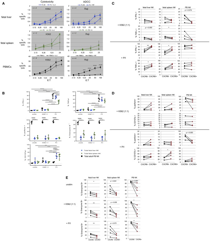
Figure 5.
Fetal liver and spleen NK cells have low killing capacity but produce cytokines and degranulate in response to stimulation. (A) Bulk fetal liver (n = 4) and spleen (n = 2) lymphocytes were plated at various effector to target cell ratios (E:T) with 51Cr-labeled target cells. K562 were used as targets for cytotoxicity assays in the presence or absence of IL-2. Raji B lymphoma cells were used as targets for ADCC, plus or minus rituximab. PBMCs from healthy donors were used as controls. (B) Intracellular flow cytometry (ICFC) was used to assess fetal liver and spleen NK cell function. Total NK cells were gated for markers of cytotoxic function including production of IFNγ, TNFα, perforin, Granzyme B, and CD107a (degranulation). Total NK cells were stimulated with CFSE-labeled K562 target cells at a 1:1 ratio or PMA/ionomycin (P/I). Unstimulated cells (unstim) were incubated in media alone. Incubation was for 4 h. Fetal liver (blue circles), fetal spleen (green circles), and PB NK cells (black squares) (n = 4). (C–E) CXCR6+ fetal liver and spleen NK cells possess a unique functional profile with respect to mediators of NK cell effector function. Differences in the contribution of CXCR6− NK cells (black circles) and CXCR6+ NK cells (red circles) in the production of IFNγ, and TNFα, (C) perforin, and CD107a (D) and Granzyme B, (E) in fetal liver and spleen NK cells following stimulation. PB NK cells were used as controls. Matched pairs were analyzed for statistical significance using the Student's t-test. P-values are as shown or as detailed in the legend to Figure 1.

