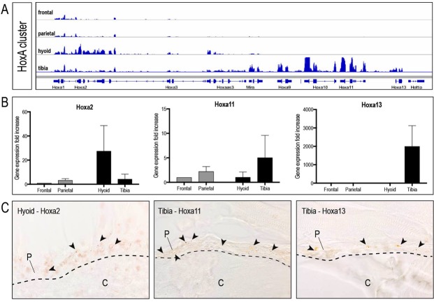
Figure 1.
Embryonic Hox status of periosteal stem/progenitor cells is preserved into adulthood. (A) Transcriptional map depicting normalized FPKM expression values for genes within the HoxA cluster. Note the near absence of Hox expression in the frontal and parietal bone, while proximal Hox genes are represented in the hyoid sample, and distal Hox genes are expressed in the tibia, similar to their embryonic pattern. Expression values from isolated periostea were averaged for each skeletal element (n = 3). (B) qPCR validation (mean +/− standard error) of three relevant Hox genes (Hoxa2, Hoxa11 and Hoxa13) identified as differentially expressed by RNA sequencing (n = 3). (C) In situ hybridization of hyoid and tibial periosteum with Hoxa2, Hoxa11 and Hoxa13 RNA antisense probes confirming spatial expression of the respective Hox genes within the periosteum (arrowheads)(n = 3). Abbreviations: c, cortical bone; p, periosteum.

