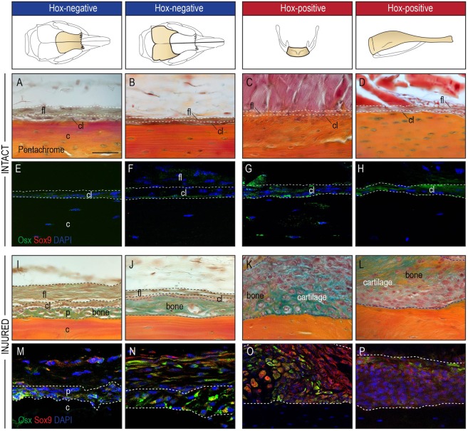
Figure 3.
Hox status imparts unique regenerative response on periosteal cells. (A–H) Representative histologic appearance of the periosteum of frontal, parietal, hyoid bone and tibia with characteristic one-cell-layer thick cambial layer (cl) containing osx-positive bone-lining cells (green cells in immunofluorescence staining)(E–H), and a thicker fibrous layer (fl) in the periphery (n = 5). (I,J) A periosteal scratch injury results in a pure osteogenic response (between dashed line) of the periosteum in the frontal and parietal bone, while (K,L) periostea from the hyoid and tibia responded with a mixed osteochondrogenic response (osx for osteogenesis, sox9 for chondrogenesis)(n = 5). (M,N) Immunofluorescence staining of the periosteal injury confirmed the osteogenic response with Osx-positive cells within the regenerate (between white dashed lines) and an absence of Sox9-expressing cells. (O,P) In contrast, the periosteal response to injury (between white dashed lines) of the hyoid and tibia exhibited both Osx+ and Sox9+ expressing cells, in line with a mixed osteochondrogenic healing response. Abbreviations: c, cortical bone; cl, cambial layer; fl, fibrous layer; p, periosteum.

