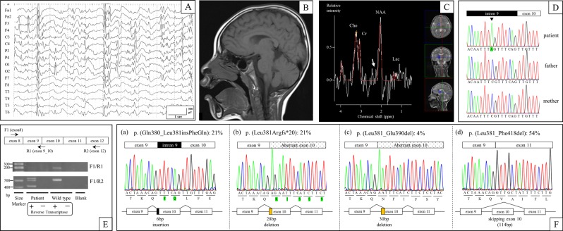
Fig. 1. Clinical findings and genetic tests of the patient.
a EEG performed at 4 years showed focal epileptic discharge with generalization in multiple foci. b Brain MRI (T1 weighted sagittal) performed at 4 years did not show any abnormal findings. c Brain MRS in the basal ganglia performed at 4 years did not show any abnormal glutamate/glutamine peaks (white arrow). d The patient carried a de novo hemizygous SLC9A6 mutation (NM_006359.2:c.1141-8C>A) that was confirmed by Sanger sequencing. e RT-PCR analysis identified multiple aberrant transcripts but no canonical transcripts in the patient, while it identified only canonical transcripts in control DNA (wild type). f: Transcript variants in the patient. Twenty-one percent of transcripts included intronic 6-bp nucleotides (a), 21% excluded exonic 28-bp nucleotides (b), 4% excluded exonic 30-bp nucleotides (c), and 54% skipped exon 10 (d)

