Figure 3.
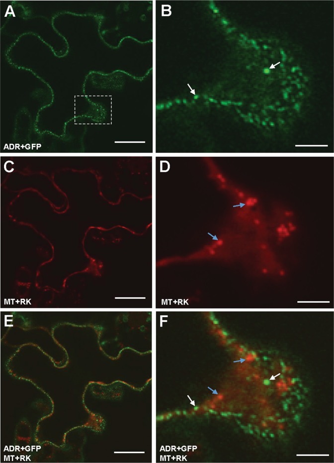
Transient expression of ADR + GFP and MT + RK in tobacco cells. (A–D) Agrobacterium-mediated transient expression of ADR + GFP (A,B) and MT + RK (C,D) in epidermal cells of N. benthamiana. ADR + GFP fusion protein accumulated in organelle-like structures (white arrows in B), whereas MT + RK was localized in mitochondria (blue arrows in D). (E,F) A merged fluorescence image of (A and C) in (E); (B and D) in (F) showing the different localization of ADR + GFP (white arrows in F) and MT + RK (blue arrows in F). (B,D and F) are close-up images (boxed) from (A,C and E), respectively. Scale bars: 20 μm in (A,C and E) and 5 μm in (B,D and F).

