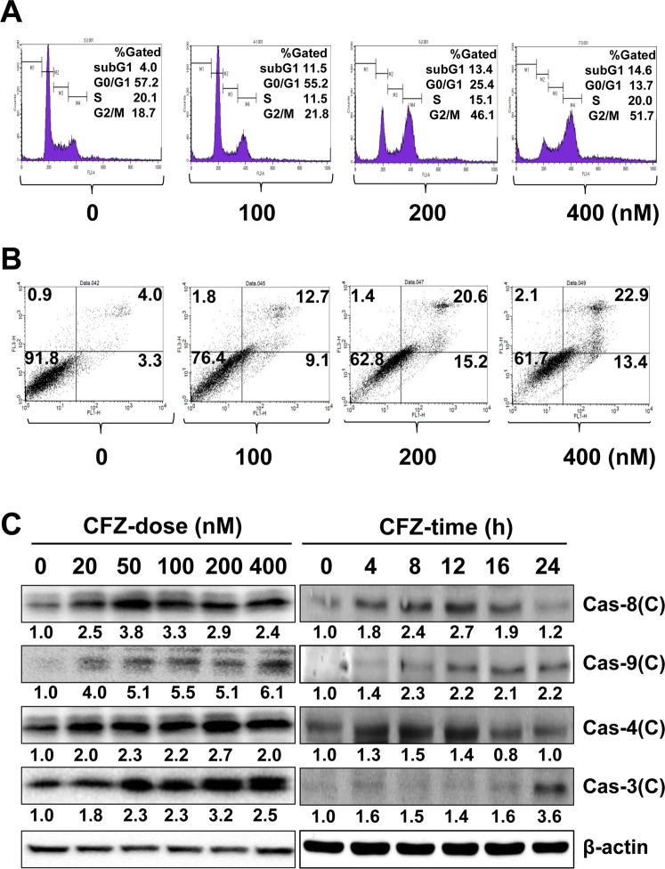
Figure 2.
Effect of CFZ on cell cycle arrest and apoptosis in SK-N-BE(2)-M17 cells. (A) SK-N-BE(2)-M17 cells were treated with vehicle or 100–400 nM CFZ for 24 h and then stained with PI and analyzed by flow cytometry. The percentage of gated cells in the subG1, G0/G1, S, and G2/M areas is shown in the upper right area of each plot. (B) Cells were treated with vehicle or 100–400 nM CFZ for 24 h. Vehicle- or CFZ-treated cells were stained with PI and annexin V-FITC and evaluated by FACS analysis. The lower right and upper right areas represent the percentages of early apoptotic cells and late apoptotic cells, respectively. The fraction of necrotic cells is shown in the upper left area. (C) Cells were treated with various concentrations (20–400 nM) of CFZ for 24 h (CFZ-dose) or treated with 200 nM CFZ for the indicated durations (CFZ-time). Cells were then lysed and total cell extracts were resolved by SDS-PAGE. The levels of activated caspases were detected by western blot analysis using antibodies against cleaved forms of caspases (Cas-8, 9, 4, and 3), and β-actin (internal control). Blots are representative of those obtained in more than three independent experiments.

