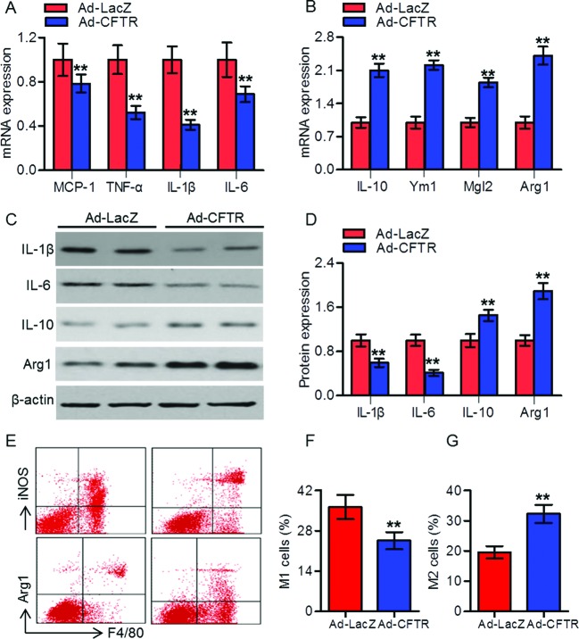
Figure 3. CFTR reduced inflammatory cytokines in aorta and peritoneal macrophages from atherosclerotic mice.
(A,B) RT-PCR analysis for the mRNA expression of MCP-1, TNF-α, IL-1β, and IL-6 (A), and IL-10, Ym1, Mgl2, and Arg1 (B) in aorta from HFD-fed apoE−/− mice treated with Ad-LacZ or Ad-CFTR. **P<0.01 compared with HFD + Ad-LacZ, n=6 in each group. (C) The protein expressions of IL-1β, IL-6, IL-10, and Arg1 in peritoneal macrophages were determined by Western blotting. (D) Quantitation of these protein expressions. **P<0.01 compared with HFD + Ad-LacZ, n=6 in each group. (E) CFTR up-regulation reduced the proportion of iNOS-positive cells (M1 phenotype) and shifted the macrophage phenotype to Arg1-positive cells (M2 phenotype) compared with Ad-Lacz group. (F) Percentage of M1 macrophages. (G) Percentage of M2 macrophages. **P<0.01 compared with HFD + Ad-LacZ, n=6 in each group.

