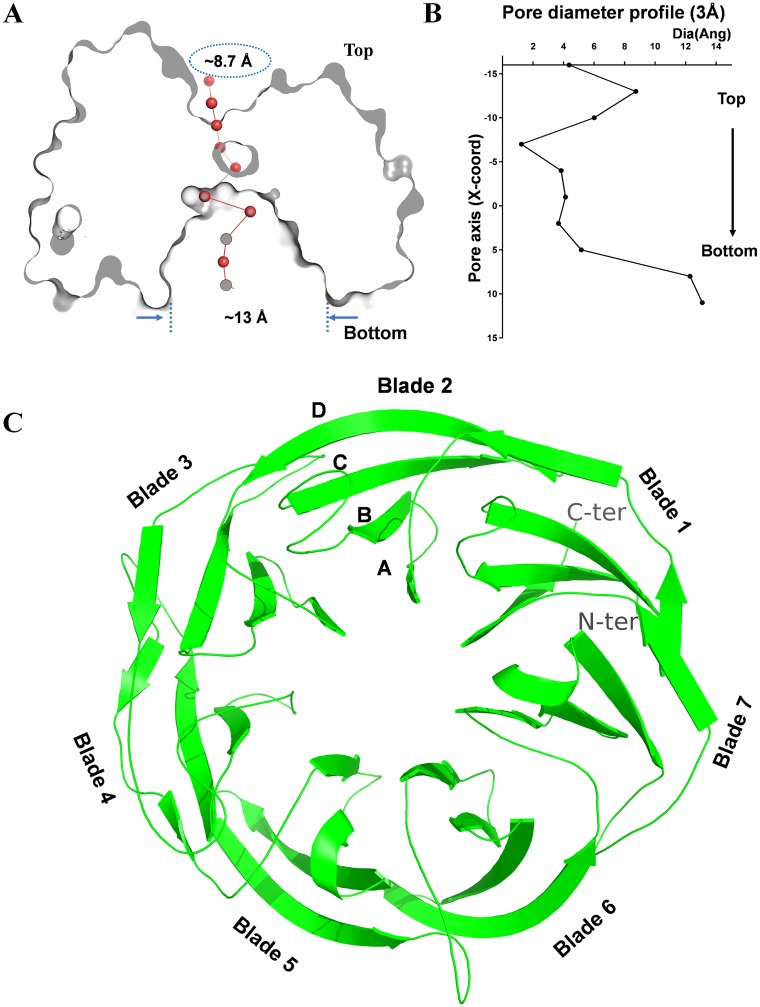
FIG 1.
Overall structure of PpgL from P. aeruginosa. (A) View rotated 90° about the horizontal axis. The structure is sliced through the center to highlight the depression and the tunnel located on the top and bottom face of PpgL, respectively. (B) Pore diameter profile at 3-Å steps. (C) Ribbon diagram of PpgL viewed down the central axis. As observed in the 1.65-Å structure, the blades are numbered 1 to 7 with the 3 + 1 arrangement of β strands from the first and last blades: the C-terminal strand and the N-terminal part of the 7th blade are indicated. Strands in each blade are labeled A to D.

