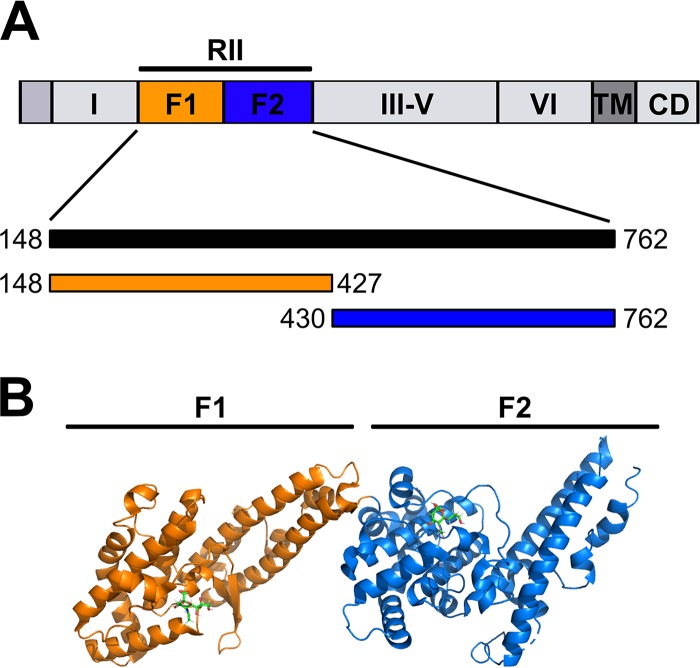
FIG 1.
Domain structure of the full-length PfEBA-140 and the details of the two DBL domains in region II. (A) EBA-140 is comprised of regions I to VI: RII consists of two DBL domains, RIII to RV contain coiled-coil like repetitions, and RVI is a conserved cysteine-rich domain. A transmembrane (TM) domain and a cytoplasmic domain (CD) are located at the carboxyl terminus. The amino acid boundaries for the RII domain construct, as well as the F1 domain and F2 domain, are shown below in the domain structure model. (B) Crystal structure of RII in complex with sialyllactose adapted from the crystal structure 4JNO. Orange, F1 domain; blue, F2 domain. Sialyllactose is represented by a stick model colored green.

