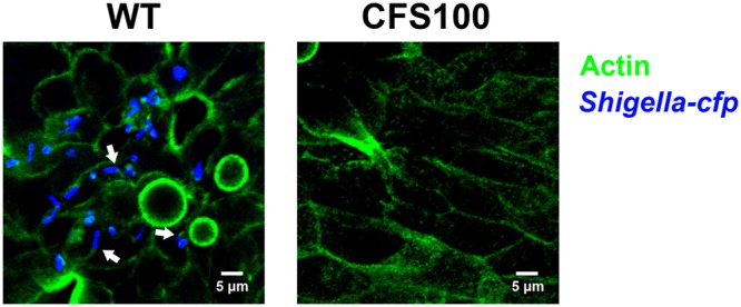
FIG 5.

Shigella forms actin tails within human colon enteroid cells. S. flexneri WT and CFS100 strains expressing cyan fluorescent protein (blue) were introduced basolaterally to enteroid monolayers grown on transwells and visualized using confocal microscopy. Actin was stained with labeled phalloidin (green). Micrographs are average-intensity projections of a z-stack. Arrows indicate intracellular Shigella bacteria associated with actin tails.
