
Figure 1.
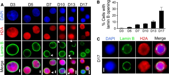
Caspase‐3‐mediated nuclear opening formation with histone release during human terminal erythroid differentiation. A, Immunofluorescent stains of lamin B and H2A in differentiating primary human erythroblasts at indicated days in culture. Arrows indicate lamin B openings. Scale bars: 5 µm. B, Quantitative analysis of the percentage of cells with lamin B opening in A. C, Immunofluorescent stains of lamin B and H2A in culture human erythroblasts on day 17. Scale bars: 2 µm