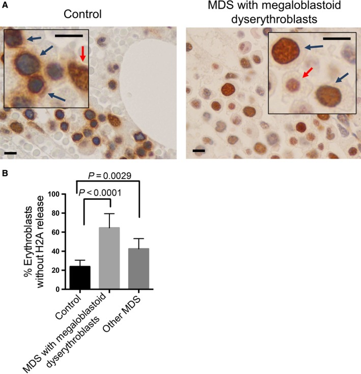
Figure 5.

Myelodysplastic syndrome (MDS) patients exhibit reduced nuclear opening formation and histone release in erythroblasts. A, Representative immunohistochemical stains of H2A in normal control individuals and patients with myelodysplastic syndromes carrying megaloblastoid erythroblasts. Blue arrows indicate erythroblasts. Red arrows indicate other hematopoietic cells. Scale bars: 8 μm. The unstained cells are red blood cells due to hemorrhage during biopsy. B, Quantitative analysis of the percentage of erythroblasts without H2A release. Control: N = 6, MDS with megaloblastoid erythroblasts: N = 10, Other MDS: N = 7. 500 erythroblasts were counted in each case
