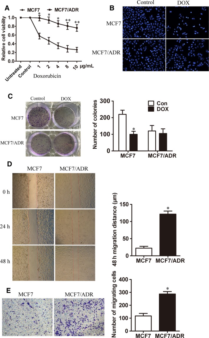
Figure 2.

DOX‐resistant cells MCF7/ADR exhibited enhancive migratory phenotype. (A) The chemo‐sensitivity of MCF7 and MCF7/ADR cells to different concentrations of DOX for 48 h treatment was evaluated by CCK8 assay (n = 3, **P < 0.01, *P < 0.05). (B) The nuclei of MCF7 and MCF7/ADR cells were stained by DAPI after treated with 4 μg/mL DOX for 48 h. (C) Colony formation was performed to detect the growth of MCF7 and MCF7/ADR cells treated with 4 μg/mL DOX (n = 3, *P < 0.05). (D) The migration ability of MCF7 and MCF7/ADR cells was measured by cell scratch test (Bar = 750 μm, n = 3, *P < 0.05). (E) Transwell migration assay was used to detect the number of trans‐membrane cells (Bar = 500 μm, n = 3, *P < 0.05)
