
Figure 4.
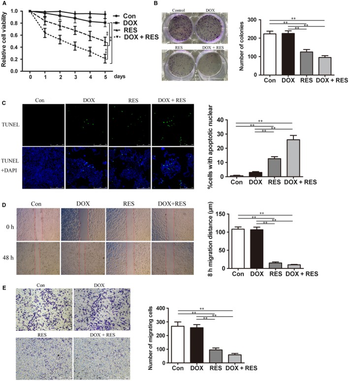
RES combined with DOX effectively inhibited proliferation and migration of MCF7/ADR cells. (A) MCF7/ADR cells were untreated or treated with 4 μg/mL DOX, 50 μmol L−1 RES, or both for 7 days, respectively and the cytotoxicity was detected by CCK8 assay (n = 3, **P < 0.01). (B) Colony forming ability of MCF7/ADR cells after being exposed to DOX or/and RES for 48 h was investigated by colony‐forming assay (n = 3, **P < 0.01). (C) MCF7/ADR cells were untreated or treated with DOX or/and RES for 48 h, followed by TUNEL analysis to detect cell apoptosis. The green fluorescence represented apoptotic bodies (n = 3, **P < 0.01). (D) The migration distance was measured to show the migration ability of MCF7/ADR cells after untreated or treated with DOX or/and RES for 48 h (Bar = 750 μm, n = 3, **P < 0.01). (E) MCF7/ADR cells were untreated or treated with DOX or/and RES for 48 h, followed by transwell migration assay was also used to detect cell migration (Bar = 500 μm, n = 3, **P < 0.01)