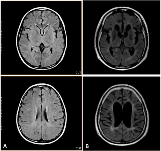
Fig. 2.

(A) Initial brain magnetic resonance imaging following patient diagnosis. Axial fluidattenuated inversion recovery (FLAIR) images were unchanged. (B) Eight years following diagnosis and treatment of subacute sclerosing panencephalitis with interferon-α, periventricular signal changes and diffuse brain atrophy were observed in these axial FLAIR images.
