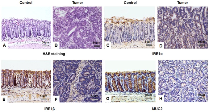
Figure 2.
IHC staining suggests that AOM/DSS treatment affects IRE1α, IRE1β and MUC2 expression in tissues. Following a one-off treatment with AOM and three cycles of DSS, tumor tissues from mice in the tumor group (n=10) and colon tissues from mice in the untreated control group (n=10) assessed by IHC. Hematoxylin and eosin staining of (A) control and (B) tumor tissues identifying typical tumor characteristics, including disordered mucosa, destroyed crypt structure, less differentiated cells, a larger cell and nuclei size, and an increased nucleus-to-cytoplasm ratio. IRE1α protein was stained using IHC in (C) control and (D) tumor tissues and expression in the cytoplasm of colon submucosa cells was observed. IHC visualized IRE1β protein in (E) control and (F) tumor tissues and identified cytoplasmic expression in colonic mucosa epithelial cells. MUC2 protein expression was visualized using IHC in goblet cells of (G) control and (H) tumor tissues. All images were obtained using a Nikon Ds-Fi2 500w [Ds-Fi2; light microscope (magnification, ×200)]; scale bar, 50 µm. IHC, immunohistochemistry; AOM, azoxymethane; DSS, dextran sulfate sodium; IRE, inositol-requiring enzyme; MUC, mucin.

