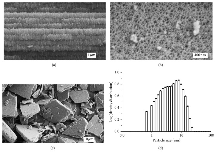
Figure 2.
(a)–(c) FESEM images produced of the mesoporous THCPSi structure of the inactive reference substrates. (a) A cross-sectional view of the layer-like structure of the mesoporous surface. Each layer has an approximate thickness of 200 nm. (b) Image taken from above the structure depicts the inlets of the pores from the top. (c) Image produced of the mesoporous THCPSi particle distribution after ultrasonication. (d) Size distribution of the THCPSi reference particles obtained by laser diffraction.

