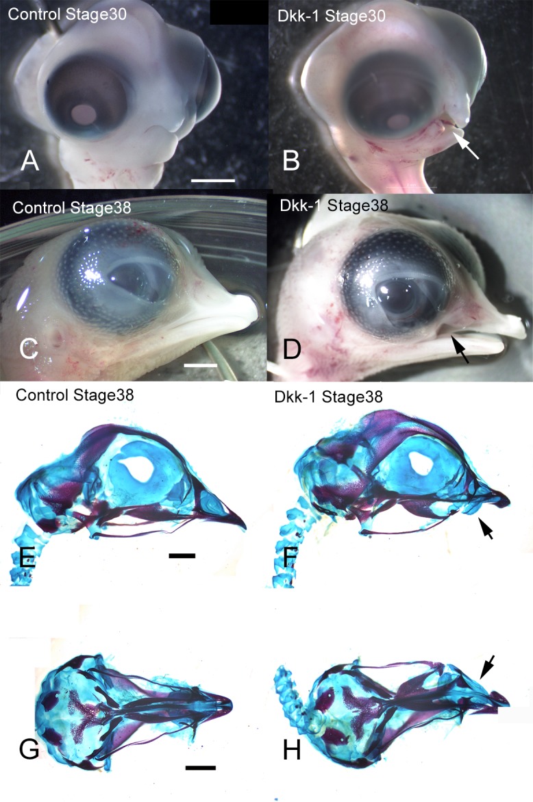
Fig. 1.

Skeletal and external changes of chick face after Dkk-1-soaked bead implantation. Beads were implanted into the right side of the maxillary prominences at stage 22 and the embryos were fixed at stage 30 or stage 38 (A, B, C, D). Control embryos (A, C) were treated with 2% BSA-soaked beads. Dkk-1-soaked beads caused a defect of the maxilla at stage 30 (B, white arrow) and stage 38 (D, black arrow). Lateral (E, F) or axial (G, H) views of heads visualized with the skeletal stains Alcian blue and Alizarin red are shown at stage 38. Hypoplasia of the premaxilla and palatine bone occurred (black arrows in (F) and (H), respectively). Bars = 5 mm.
