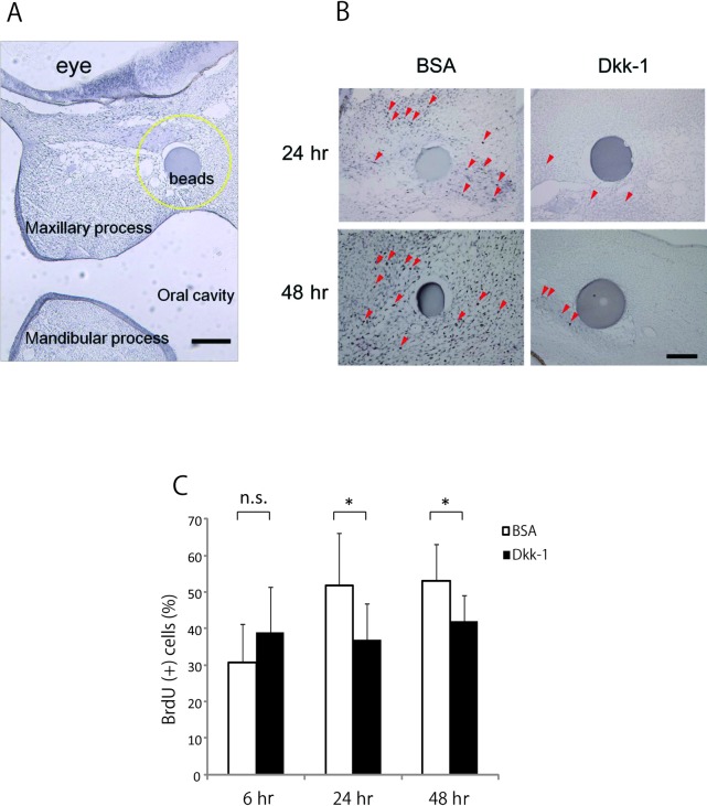
Fig. 2.
BrdU labeling and immunohistochemical staining to evaluate cell proliferation. (A) The area in which BrdU-positive cells were counted around an implanted bead (within a radius of 100 μm) is indicated by a yellow circular line. (B) BrdU-labeled cells (red arrowheads) were more abundant around BSA-treated beads (n = 5) than around Dkk-1-treated beads (n = 5). Bars = 100 μm. (C) The statistical results of BrdU-positive cells in the maxillary prominences treated with BSA and Dkk-1 after 6 hr, 24 hr, and 48 hr. Values are the mean ± S.D. in separate experiments using 9 embryos. *: P < 0.05.

