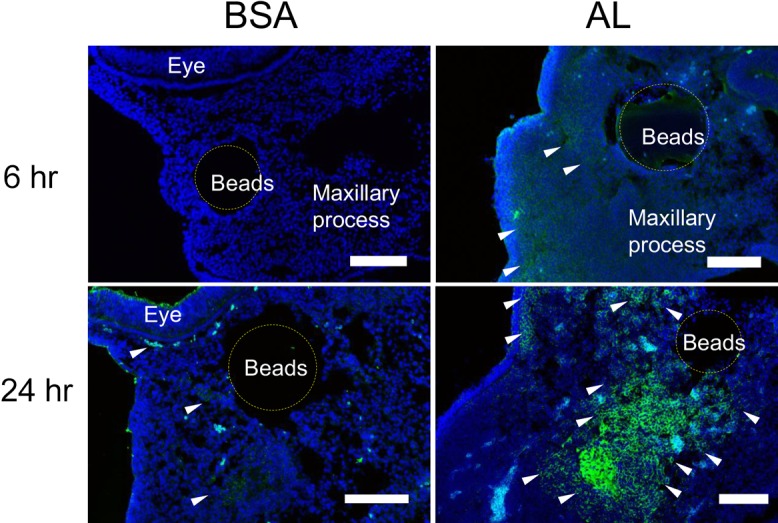
Fig. 5.

Expression of N-cadherin in the maxillary prominences of embryos at 6 hr and 24 hr after treatment. N-cadherin immunolabelings (green, white arrowhead) were expressed in the extracellular space around the beads. Nuclei were counterstained with DAPI (blue). The N-cadherin labeling increased following AL-soaked bead implantation in the maxillary prominences at 24 hr after treatment (n = 3/3). Bar = 100 μm.
