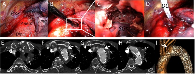
Fig. 2 Video-assisted thoracoscopic debridement for delayed abscess after thoracic endovascular aortic repair.
(A–D) Thoracoscopic findings at the time of the debridement. (A) Overview of the aneurysm located on the aortic arch. (B) The aneurysm was partially opened at the middle (white square). (C) Enlarged view inside the aneurysm. Infected tissue around the stent-graft (SG) was sufficiently excised, and thus the stent-graft was partially exposed. (D) A drainage catheter (DC) was placed in the cavity of the aneurysm after the debridement. (E–I) Computed tomography following the debridement. A delayed pseudoaneurysm appeared at the proximal edge of the stent-graft (large arrows). Small arrows: drainage catheter.

