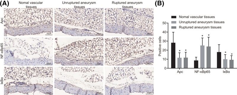
Figure 1. Down-regulation of Apc and IκBα and up-regulation of NF-κB p65 in unruptured and ruptured IA tissues were revealed by immunohistochemstry (× 400).
(A) Immunohistochemical staining for protein Apc, NF-κB p65, and IκBα in normal vascular tissues, unruptured and ruptured IA tissues. (B) Quantitative analysis for positive expression rate; *P<0.05 compared with normal vascular tissues.

