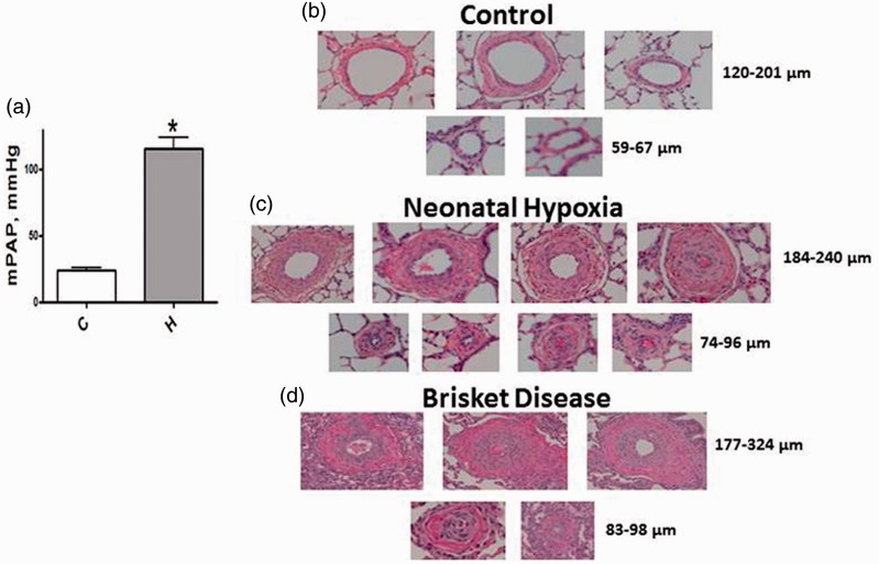
Fig. 2.
(a) A significant increase in mPAP is seen in neonatal calves exposed to hypobaric hypoxia (n = 7–9). C, controls; H, hypoxic group. *P < 0.05 vs. controls. (b) Thin walled pulmonary arteries of varying sizes from normal neonatal calves. (c) Arteries from neonatal hypoxia groups show significant medial thickness and lumens in some arteries appear obliterated. (d) Pulmonary arteries from steers with Brisket disease also show significant medial wall thickening and occlusion of some arteries.

