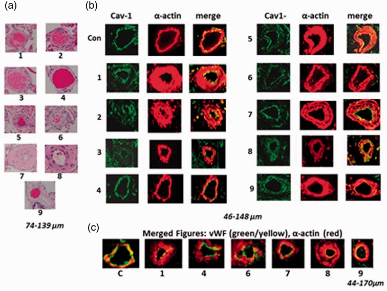
Fig. 8.
(a) H&E staining of the pulmonary arteries from Patients 1–9. Most arteries show varying degrees of medial thickening except Patient 4, despite extensive emphysema. (b) Immunofluorescence study depicting the expression of cav-1 (green) and smooth muscle α-actin (red) in pulmonary arteries. The control artery (Con) and the arteries from patients with chronic lung display the presence of endothelial cav-1. (c) Representative immunofluorescence study showing the expression of vWF (yellow/green) and smooth muscle α-actin (red) as merged figures. There is no loss of vWF in the group of patients with the lung diseases we studied.

