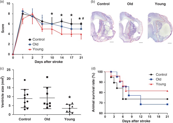
Figure 3.
Assessment of outcomes and brain atrophy with Cresyl violet staining after treatment. (a) Rats treated with young human mesenchymal stem cells (hMSCs) (n = 9) showed better behavioural recovery by modified neurological severity score than controls (n = 10) or old hMSCs (n = 8) (Steel–Dwass test, *P < 0.05 vs. Control, #P < 0.05 vs. Old hMSC). Each point represents the median (interquartile range, 25–75th percentile) in the graph. (b) Cresyl violet staining. Scale bar = 1 mm. (c) Left ventricle size in young hMSCs was significantly smaller than controls or old hMSCs (one-way ANOVA, Tukey–Kramer multiple comparison test, *P < 0.05). Data are presented as mean ± SD. (d) There were no significant differences in animal survival rates between the three groups (n = 26 in young hMSC group, n = 25 in old hMSC group and n = 19 in controls; Log-rank test).

