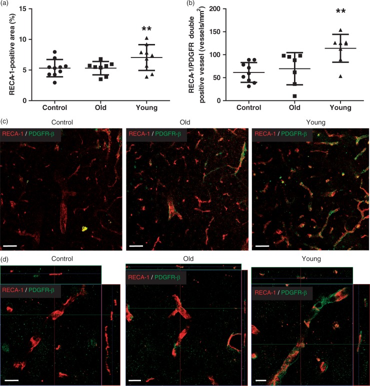
Figure 6.
Analysis of angiogenesis in the peri-infarct cortex at D21. (a) Immunohistochemical staining at the peri-infarct cortex showed that the RECA-1-positive area in the young human mesenchymal stem cell (hMSC) group was significantly larger than in controls or the old hMSC group. (b) The number of platelet-derived growth factor receptor (PDGFR)-β-positive vessels in the young hMSC group was higher than in controls or the old hMSC group (one-way ANOVA, Tukey–Kramer multiple comparison test, **P < 0.01). (c), (d) RECA-1 and PDGFR-β double staining images at the peri-infarct cortex. Data are presented as mean ± SD. Scale bar = 50 µm (c), 10 µm (d), magnified image.

