Figure 7.
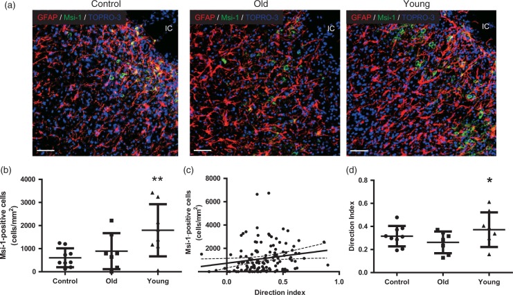
Analysis of neurogenesis in peri-infarct cortex at D21. (a) Glial fibrillary acidic protein (GFAP) and Musashi-1 (Msi-1) double staining. Scale bar = 50 µm. IC: infarct core. (b) The number of Msi-1-positive neural stem cells at the peri-infarct cortex at D21 was significantly larger in the young human mesenchymal stem cells (hMSC) group than in controls and the old hMSC group (one-way ANOVA, Tukey–Kramer multiple comparison test, **P < 0.01). (c) The direction index was significantly correlated with the number of Msi-1-positive cells migrating close to the infarct core (R = 0.2683 and P < 0.01, Spearman). (d) The direction index of the young hMSC group was significantly higher than in the old hMSC group (one-way ANOVA, Tukey–Kramer multiple comparison test, *P < 0.05). Data are presented as mean ± SD.

