Fig. 1.
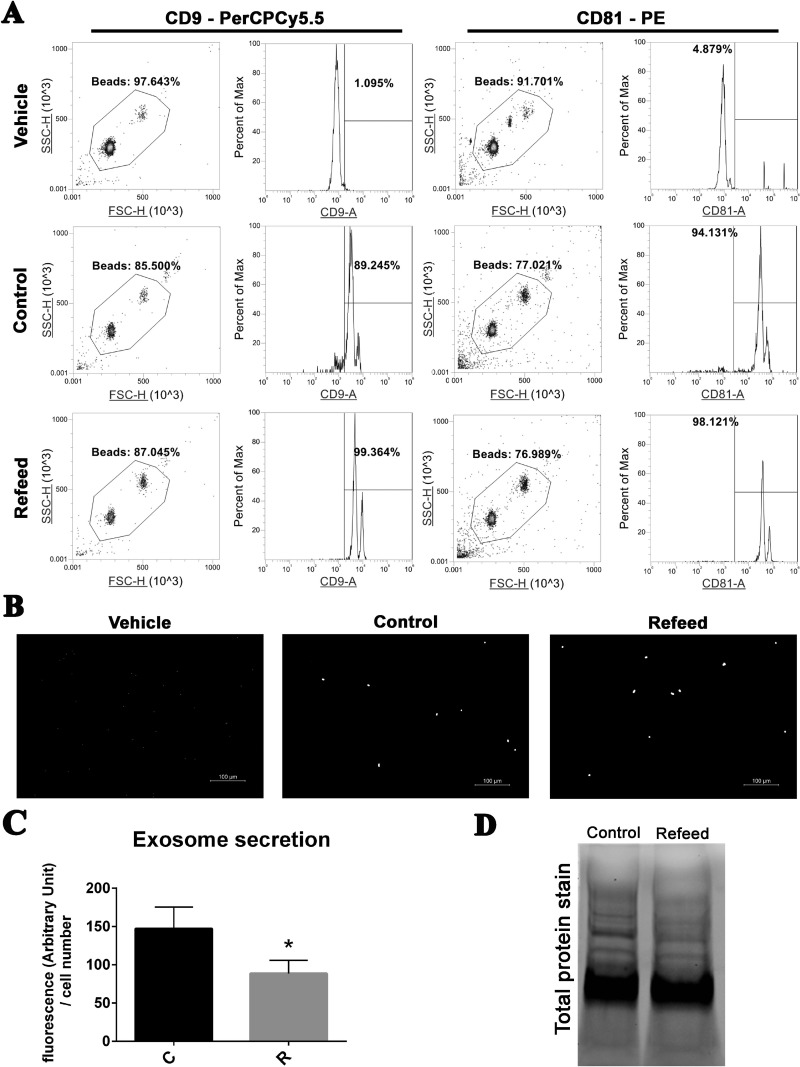
Exosome characterization and quantification in media of cells cultured with or without Refeed®. (A) Characterization of specific tetraspanin membrane proteins CD9 and CD81 by cytofluorimetric analysis in CD81-enriched exosomes derived from control (Control) or Refeed®-supplemented (Refeed®) cells; vehicle alone (Vehicle) analysis is also shown. Density plots showing gating strategy used to sort exosomes-anti-CD81 bead complexes (left panel). Histogram plots showing fluorescence intensity detected for CD9 or CD81 from the gated beads (right panel). The numbers above the gating bars on the histograms depict percentage of positive gated beads for the selected marker. Representative of 3 different analysis. (B) Representative wide-field fluorescence gray-scale images of exosomes-bead complexes stained with anti-CD81 PE antibody. (C) Quantification of exosomes stained with BODIPY® TR ceramide isolated from control (C) or Refeed®-supplemented (R) human fetal membranes MSCs. Fluorescence values were normalized on cell number of each sample. *Significantly different from control-derived exosomes (n = 6, statistical test: 2-tailed, unpaired Student’s t-test, *p < .05). (D) Stain-free acquisition of total protein content of control and Refeed® cell-derived exosomes. Representative of 3 different analyses.

