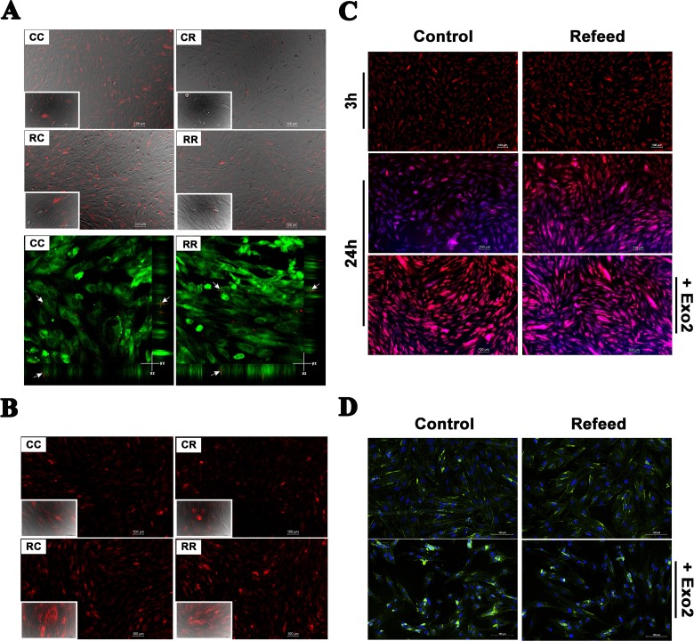
Fig. 3.
Vesicle trafficking is modified by Refeed® supplementation. Control and Refeed® cell-derived exosomes were added to culture medium of control and Refeed®-supplemented hFM-MSCs. (A) Three hours after exosome supplementation, acquisitions were performed. Live imaging merge of Differential Interference Contrast (DIC) and fluorescence channel of control or Refeed®-supplemented hFM-MSCs incubated with PKH26-labeled exosomes (red spots; derived from control [CC or RC] or Refeed®-supplemented cells [CR or RR]). Images confirm quantification data, as control exosomes appear more numerous in every captured field. Confocal live images (lower panel) at 20 min confirmed exosome internalization into cytoplasm of cells counterstained with PKH2 (green). The arrows highlight exosome x- and y-position projection on the z-stack. No differences in terms of uptake kinetic was noticeable. (B) Twenty-four hours after exosome adding, images show different fluorescent levels, which is cell dependent and not exosome origin dependent. Fluorescence live images of control or Refeed®-supplemented hFM-MSCs incubated with BODIPY® TR ceramide-labeled exosomes (red; derived from control [CC or RC] or Refeed®-supplemented cells [CR or RR]; inset: merge of bright field with fluorescence images at higher magnification [40×]; scale bar: 50 µm). (C) Fluorescence live images of control and Refeed®-supplemented cells at 3 and 24 h after staining with BODIPY TR ceramide alone (red). ER-TrackerTM Blue-White DPX (blue) was added just prior to acquisition. Cells stained with BODIPY TR ceramide treated with Exo2 (lower panels). Images show the different turnover of BODIPY TR ceramide dye at 24 h, with a higher clearance in control cells. This difference drops in presence of the inhibitor. (D) Fluorescence merged images of control and Refeed®-supplemented cells treated (lower panels) or not (upper panels) with Exo2. CD81 (green) expression in untreated control cells and Refeed®-supplemented ones is comparable. Exo2 treatment deeply affected control cells that accumulated CD81 in large intracellular compartments. CD81 accumulation is also visible in Refeed®-supplemented cells but to a lesser extent. Cell nuclei were counterstained with NucBlue® Fixed Cell ReadyProbes® Reagent (blue).

