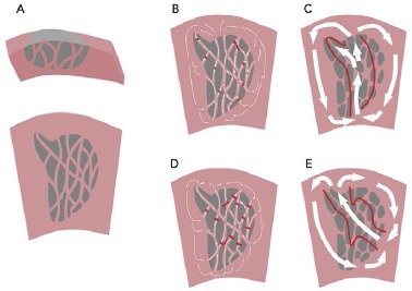
Figure 6: A Critical Zone Anchoring Multiple Ventricular Tachycardias.

A: Many years after MI, there is an area of scar extending from the endocardium. There is a fine mesh of surviving fibres on the endocardial surface. B: In ventricular tachycardia (VT), there is slow conduction, collision and functional block to create a protected isthmus channel (red lines). C: Current high-resolution technologies do not capture the complex zig-zag activation in the scar but can identify the path of activation through the critical isthmus. D: A second VT in the same scar is characterised by collision and functional block at different sites, resulting in a different pattern of activation and breakout site. E: Mapped with a high-resolution electroanatomical system, VT2 appears to have a different isthmus, which overlaps with that mapped in VT1.
