


Figure 2: A minigene containing exons 9–12 generates circ12→10.
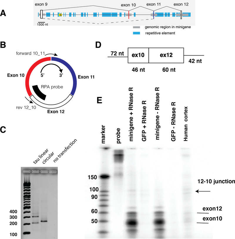
A. Structure of the minigene. Exons are indicated as vertical boxes, horizontal gray boxes indicate the genomic regions used for cloning the minigene. Blue boxes indicate repetitive elements defined by the UCSC genome browser repeat masker. The yellow box with an ‘S’ depicts the intronless Saitohin reading frame. The drawing is to scale, as indicated.B. Detection of circ12→10. The orientation of the circular RNA is clockwise 5’→3’. The location of the detection primer for10_11 and rev12_10 is indicated. The probe used for RPA is shown as a bold line.C. Detection of RNAs made from the exon 9–12 minigene. 1 μg of the minigene was transfected into HEK293 cells and after 24 hrs, RNAs were detected by RT-PCR. The negative control are untransfected HEK293 cells, using circRNA primers.D. RNase protection probe to detect circ12→10 RNA. The T7 antisense RNA is shown with the nucleotide lengths indicated., E. RNAse protection using RNA from transfected cells as well as human cortex. 50 μg total RNA was used, which was digested with RNaseR, as indicated.