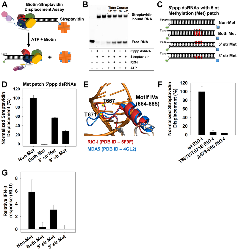
Figure 4. Directionality of RIG-I translocation and role of Helicase motif IVa.
(A) Biotin-streptavidin displacement assay schematic. (B) Time-course of biotin-streptavidin displacement from 5’ppp dsRNA (25 nM) by RIG-I (25 nM). (C) 27bp methylated patch 5’ppp dsRNAs to assess directionality of RIG-I translocation (Red bars- 5nt patch of 2’-O-methylated nucleotides placed 15bp downstream of the 5’ppp end). (D) Streptavidin displacement activity on methylated patch 5’ppp dsRNAs. Error bars are SEM from triplicates. (E) Helicase motif IVa of RIG-I (red) and MDA5 (blue). T667E and T671E in RIG-I are shown as yellow circles. (F) Streptavidin displacement by motif IVa mutants on the 5’ppp dsRNA. Error bars are SEM from triplicates. (G) IFN-β reporter activity of RIGI transfected HEK293T cells upon activation by the 2’-O-methylated 5’ppp dsRNAs. Error bars are SEM from quadruplicates. See also Figure S4.

