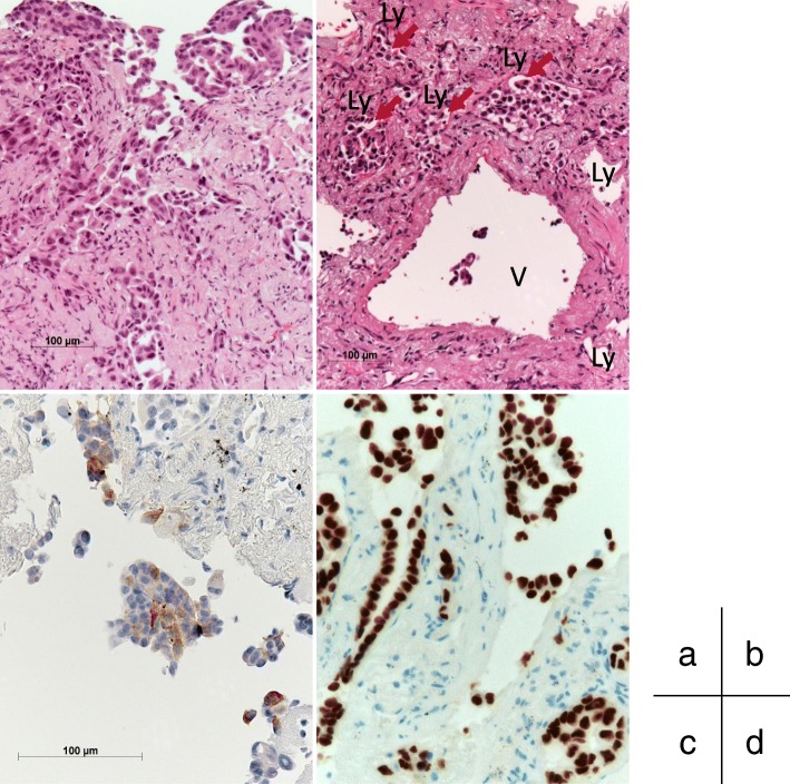
Fig. 2.
Histopathological findings of biopsy specimen from the left lung tumor (July 2016). a, b Microscopic examination showing bronchial mucosal infiltration by poorly differentiated lung adenocarcinoma (a), and vascular invasion is seen (arrows) (b) (H&E staining). c, d The cytoplasm of the tumor cells was immunohistochemically positive for surfactant protein A (c), and the tumor cell nuclei were positive for thyroid transcription factor 1 (d). Ly Lymph duct, V Vein

