Fig. 2.
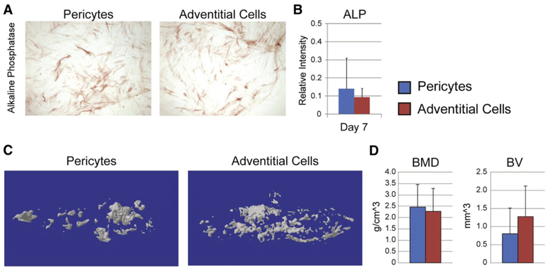
Human pericytes and adventitial cells undergo roughly similar osteogenic differentiation. (A,B) Human pericytes and adventitial cells from the same patient sample were cultured under osteogenic conditions (10% FBS, 100 μg/ml ascorbic acid, 10 mM β-glycerophosphate). (A) Representative image of alkaline phosphatase (ALP) staining at 5 d differentiation. (B) Semi-quantification of ALP staining. (C,D) Human pericytes or adventitial cells were implanted in the thigh complex of a SCID mouse using a collagen sponge carrier (2.5 × 10⁵ cells, sponge size 2.0 × 1.0 × 0.5 cm). (C) 3-Dimensional MicroCT reconstructions. (D) MicroCT based quantification of bone mineral density (BMD) and bone volume (BV).

