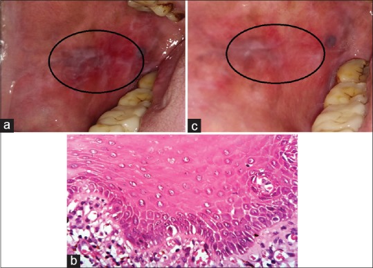
Figure 1.

(a) Pretreatment erosive lichen planus lesions on buccal mucosa of Case 1. (b) ×400 view of H and E–stained soft tissue section of Case 1 with hyperkeratotic epithelium along with underlying fibrocellular connective tissue stroma with spongiosis and acanthosis at focal areas. The underlying connective tissue is fibrocellular with dense inflammatory cells chiefly lymphocytes and cells at subepithelial connective tissue and scattered. (c) Posttreatment reduction in erosive pattern of the lesions after zinc therapy on buccal mucosa in Case 1
