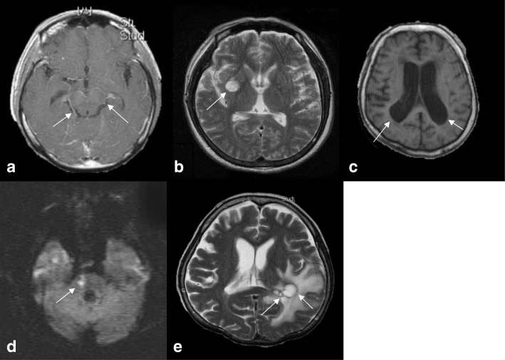
Fig. 2.
The main magnetic resonance imaging features of the elderly patients with cryptococcal meningitis. a: Gadolinium contrast-enhanced T1-weighted image (T1WI) showing basal meningeal enhancement (arrow). b: T2-weighted magnetic resonance image (T2WI) showing pseudocysts (arrow). c: T1WI showing ventricular dilatation. d: Diffusion-weighted image showing a recent cerebral infarct at the right pons (arrow). e: T2WI showing cryptococcoma (arrow)

