Fig. 3.

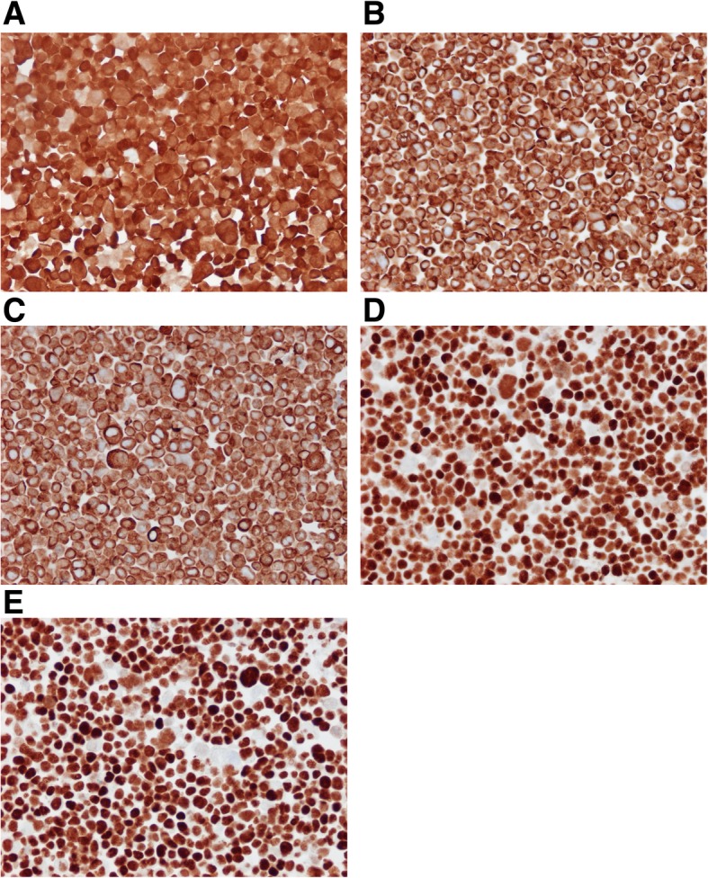
Cell pellets of LU-HNSCC-26 were sectioned in 3-μm slices and stained with antibodies against a. p16, b. CKAE1/AE3, c. CK5, d. p63 and E. p40. All photos were taken with a 20× objective

Cell pellets of LU-HNSCC-26 were sectioned in 3-μm slices and stained with antibodies against a. p16, b. CKAE1/AE3, c. CK5, d. p63 and E. p40. All photos were taken with a 20× objective