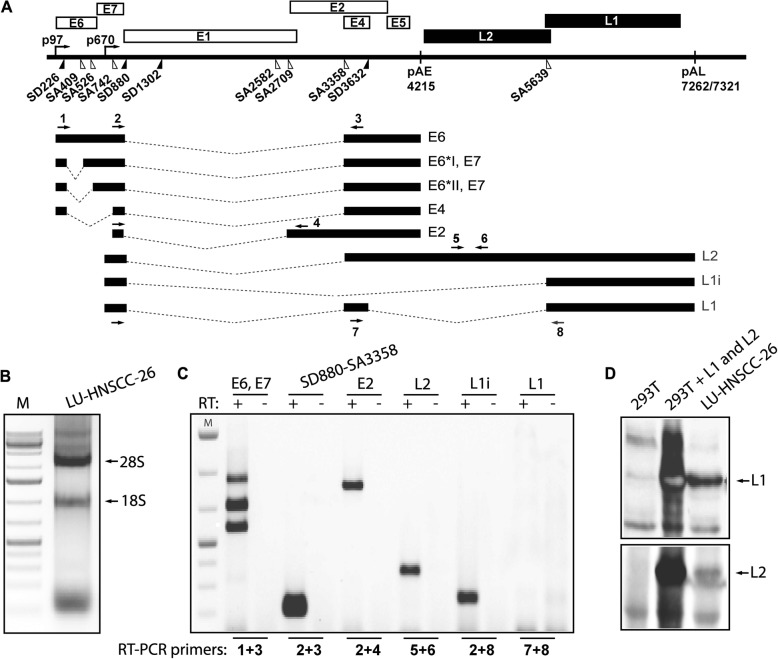
Fig. 5.
a. Schematic representation of the HPV16 genome. Rectangles represent open reading frames, promoters p97 and p670 are indicated as arrows, and filled and open triangles represent 5′- and 3′-splices sites respectively [39]. HPV16 early and late polyA signals pAE and pAL are indicated. The mRNAs produced by HPV16 cells are indicated below the genome and RT-PCR primers are indicated as arrows and numbered. RT-PCR primers are listed in Table 1. b. Total RNA extracted from LU-HNSCC-26 cells. 28S and 18S ribosomal RNAs are indicated. C. RT-PCR on total RNA extracted from the LU-HNSCC-26 cell line with the indicated primer pairs. D. Western blot with antibody against the HPV16 L1 or L2 protein on cell extracts from 293 T cells, 293 T cells transfected with a codon modified HPV16 L1 expression plasmid or LU-HNSCC-26 cells

