Figure 4.
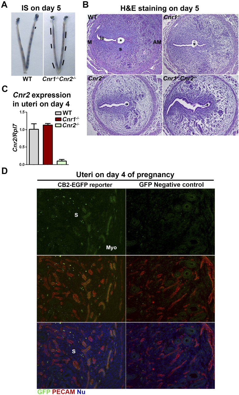
Cnr1−/−Cnr2−/− females show increased edema in implantation sites on day 5 of pregnancy. (A) Implantation sites in WT and Cnr1−/−Cnr2−/− females on day 5 of pregnancy. Lines indicate the uterine domain with blue color. (B) H&E staining in implantation sites on day 5 of pregnancy. (C) RT-PCR of Cnr2 using RNA collected from uteri on day 4 of pregnancy. Cnr2 is expressed in uterine endothelial cells. Values are means ± SEM. (D) Immunostaining of GFP and PECAM in uteri of CB2-EGFP reporter and control mice on day 4 of pregnancy. GFP-positive signals are observed on PECAM-positive endothelial cells. *Positions of embryos. AM, antimesometrial side; IS, implantation site; M, mesometrial side; Myo, myometrium; Nu, nuclear; S, stroma.

