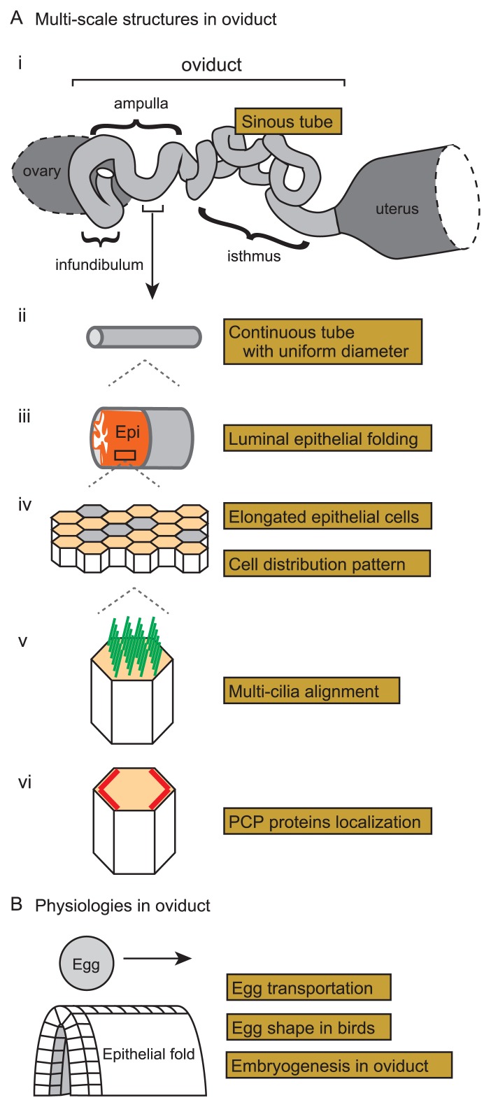
Figure 1.
Multi-scale structures and physiologies in murine oviduct. A. Multi-scale structures in the murine oviduct are illustrated. i) A whole view of the oviduct is shown with the three sub-domains: infundibulum, ampulla, and isthmus. The oviduct presents a sinuous tube when the tube is surgically disentangled. Before the surgical operation, the tube is more compactly accommodated as illustrated in Figure 7A. ii) Each sub-domain of the oviduct is a continuous tube with almost uniform diameter. iii) Multiple folds of the luminal epithelia in the infundibulum and ampulla are shown. The epithelial sheet and the surrounding smooth muscle layer are shown by orange and gray, respectively. iv) Cell distribution in the luminal epithelium is shown. The epithelial cells are comprised of ciliated (orange polygons) and secretory (gray polygons) cells, and the formers are elongated along the longitudinal axis of the tube. v) Well-aligned multiple cilia (green lines) in a ciliated cell are shown. vi) Planar cell polarity (PCP) in the luminal epithelium is described. PCP-related proteins (red lines) are localized on specific boundaries of the epithelial cells. B. Physiologies of the mammalian and avian oviducts are illustrated. In the oviducts, eggs are transported along the epithelial folds. Epithelial cells are illustrated as cubic blocks in the fold. Avian egg shapes are determined in the oviduct, which is related to the formation of the anterior-posterior axes of the embryos in the eggs.

