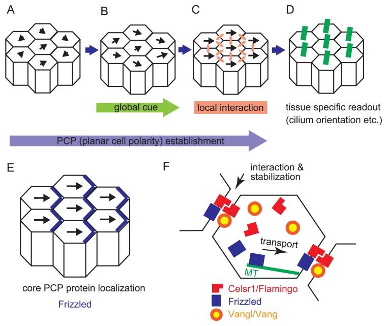
Figure 2.
Formation of planar cell polarity. A–D. Establishment of planar cell polarity (PCP) is illustrated according to the three-tiered model. Epithelial cells are shown by polygons, and their polarities are described by black arrows. The lengths of the arrows mean the strengths of the polarities. A. Before the establishment, epithelial cells have weak polarities with randomized directions. B. The polarities in the epithelial cells in the presence of a global directional cue are shown. C. The polarities in the presence of local signal propagation are shown. Between adjacent cells, local signals are transduced (light red arrows). Consequently, all epithelial cells exhibit almost the same direction of the polarities. D. Tissue specific readout of PCP established in C includes cilium orientation etc. Cilia are exemplified. They are described by green lines, and orient toward right sides of the cells in this panel. E. A relationship between the directions of the cellular polarities and localization of core PCP proteins is illustrated. The localization of Frizzled proteins is exemplified (blue lines). F. Molecular mechanisms of PCP establishment are described. The epithelial cells are shown by polygons. Several core PCP proteins are shown by red, blue, and orange/yellow. These proteins are located on specific boundaries of the cells. On the cellular boundaries, these proteins might form complexes with proteins from neighboring cells, which contributes to the stabilization of the localization. Some of core PCP proteins are transported to the specific boundaries along microtubules (green lines). In the oviduct, the Celsr1 proteins are localized on the boundaries of both the ovary and the uterus sides, whereas the Vangl proteins are localized on the boundaries of the ovary side.

