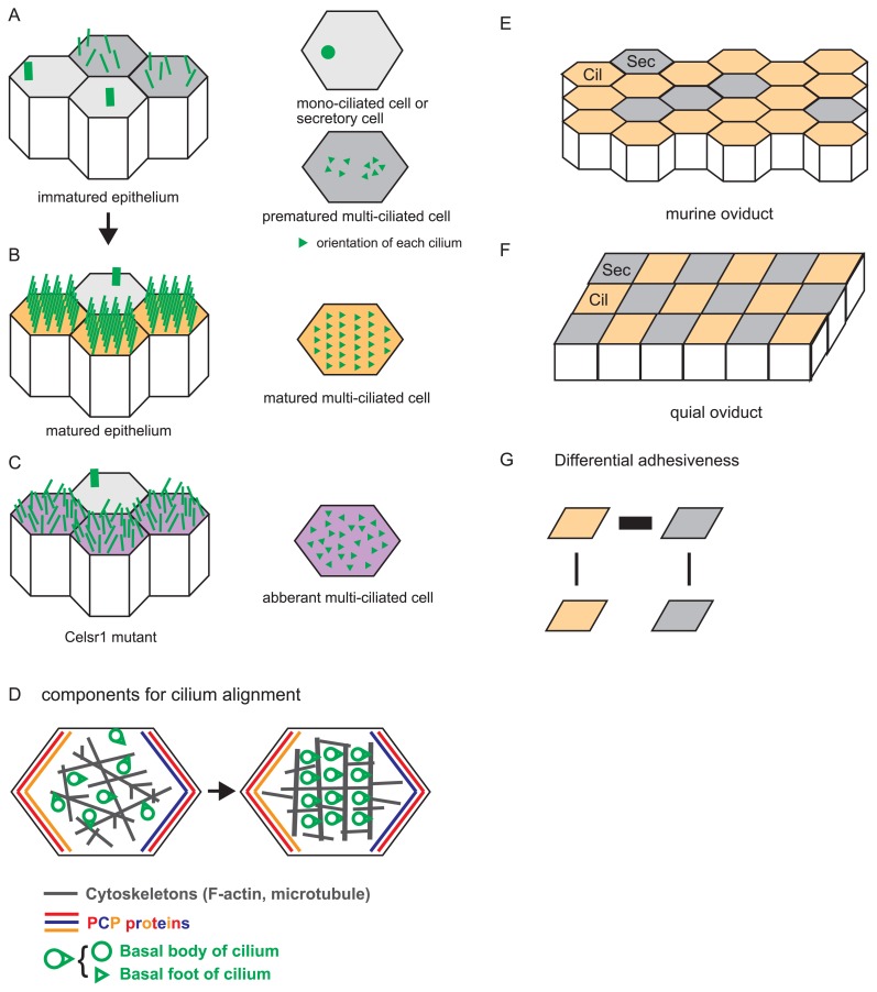
Figure 3.
Alignment of multiple cilia in a single cell and a tissue, and distribution of ciliated cells. A. An immature epithelium is illustrated with cilia. Cells are depicted as polygons, and each cilium is shown by a green line (left panel). There are mono-ciliated cells and premature multi-ciliated cells as shown in the right panel where cilia are described as green triangles whose orientations are randomized. B. A mature epithelium is illustrated with cilia. The cilia are ordered, and their orientations are aligned in multi-ciliated cells. Mono-ciliated cells are also observed. C. A mature epithelium in the Celsr1 mutant mice is illustrated with cilia. The cilia are not ordered, and their orientations are not aligned in multi-ciliated cells. D. Components involved in cilium alignment and the process of cilium alignment are described. A single epithelial cell is shown by a polygon. Basal bodies and basal foots of cilia are depicted by green circles and triangles, respectively. Cytoskeletons are shown by gray. Localization of the core PCP proteins are shown by red, blue, and orange. Cilia become aligned with the cooperation of cytoskeletal filaments (from left to right panel). E. A murine oviduct epithelium is shown. Ciliated and secretory cells are described by orange polygons (Cil) and gray polygons (Sec), respectively. Three secretory cells align linearly in this panel. F. A quail epithelium is shown. Ciliated (Cil) and secretory (Sec) cells form a checkerboard pattern. G. Differential adhesion hypothesis (DAH). According to DAH, the checkerboard pattern in the quail oviducts is explained by differential adhesiveness between each pair of the cells (Cil and Sec). Thicknesses of black lines connecting the cells mean the strengths of cell-cell adhesion.

