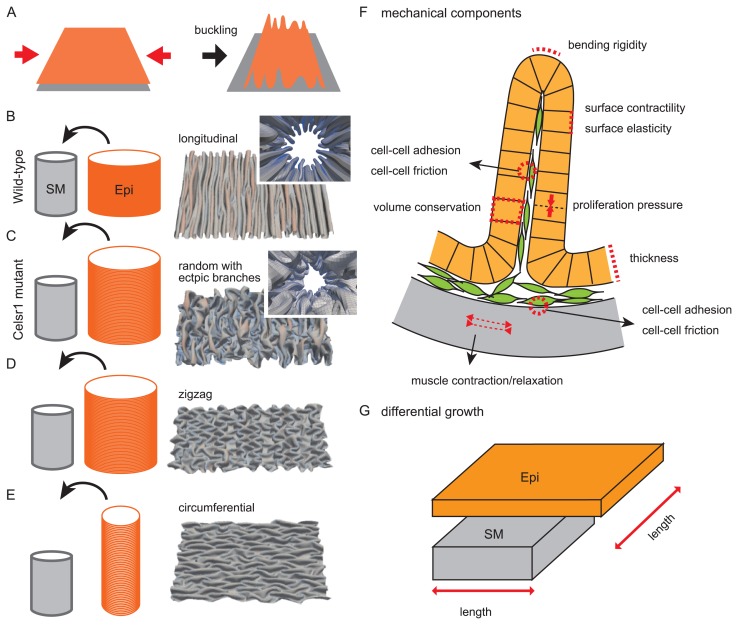
Figure 4.
Pattern formation of multiple folds. A. Buckling is explained. When an elastic sheet such as an epithelium (orange) is pushed inward (red arrows), folds are formed. B. Folding in wild-type situations is illustrated. A cylindrical epithelial sheet (orange; Epi) is put into a cylindrical smooth muscle layer (gray, SM). The circumferential length of the Epi is longer than that of the SM, whereas the longitudinal length is comparable. A luminal view with folds generated in our simulation is shown in the inset of the right panel. An opened view of the luminal side is also shown where longitudinal multiple folds are observed. C. Folding in Celsr1 mutant situations is illustrated. In contrast to the wild-type situation (B), the longitudinal length of the Epi is longer than that of the SM. The generated folds exhibit randomized directions with ectopic branches. D. Generation of zigzag folds is explained. The lengths of the Epi and SM are similar to those in C, but the outcomes can be different due to the initial condition of the simulations. E. Generation of circumferential folds is explained. The circumferential lengths of the two layers are comparable, whereas the longitudinal length of the Epi is longer than that of the SM. F. Mechanical components potentially related to fold patterns are described. A circumferential section of the oviduct is shown. An epithelial sheet (orange) forming a fold is shown with each cell (orange box). A smooth muscle layer is shown by gray. Mesenchymal cells are also shown by green. Many potential components can be considered, but we implemented only several components in our simulation explaining the fold patterns [12]. G. Differential growth or length between an epithelial sheet (Epi) and a smooth muscle layer is described. The simulation results (B–E) are cited from our previous work [12].

