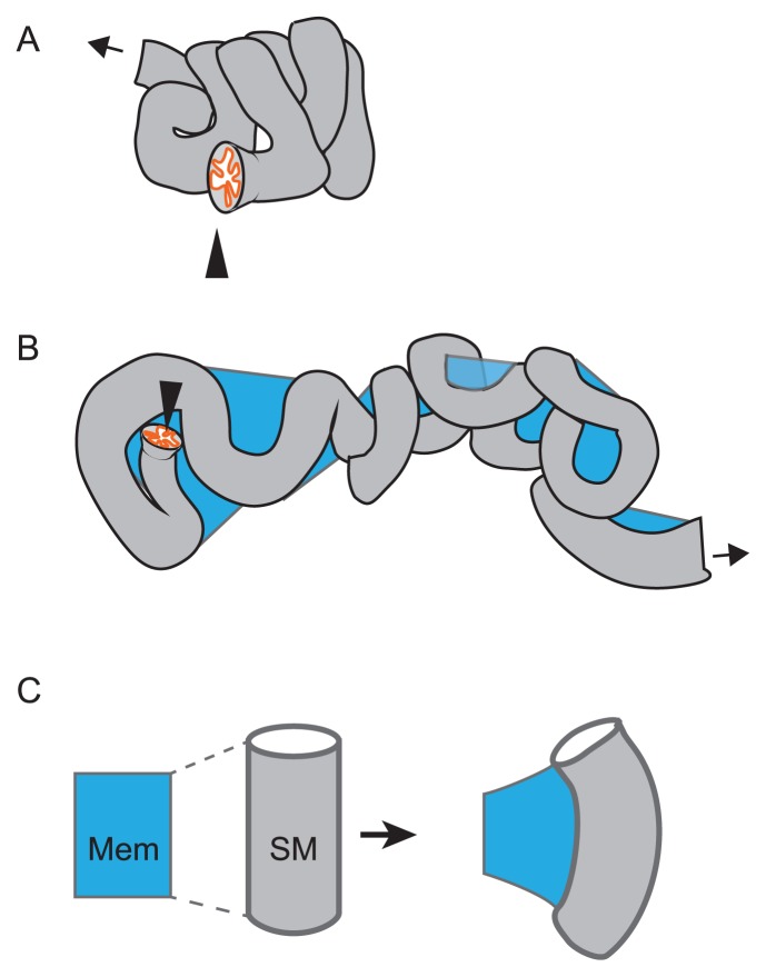
Figure 7.
Sinuous shape of tubular organ. A. A compactly accommodated murine oviduct is illustrated. The entry from the ovary is shown by an arrowhead, and the exit to the uterus is shown by an arrow. B. An oviduct surgically disentangled is illustrated. A membrane (mesosalpinx; blue) is connecting to the tube. The tube exhibits a sinuous pattern. C. Differential growth between the tube and the membrane is explained. The membrane (blue) is connecting with the smooth muscle (SM) of the tube along the longitudinal direction of the tube. If the longitudinal length of the tube is longer than that of the membrane, the tube is deformed and possibly to form a sinuous pattern described in B.

