Figure 1.
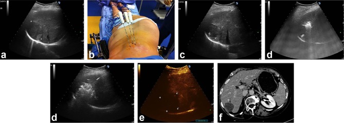
A 5.5 cm HCC tumour (between markers) is displayed in the seventh hepatic segment at US intercostal scan in a 77-year-old female with HCV-positive hepatic cirrhosis (a). Three RF needles have been sequentially positioned in a triangular configuration through two adjacent intercostal spaces (b). Ultrasound counterpart: two electrodes (4 cm exposed tip) are seen in the same US plane (needle tips identified by the markers) spaced 2 cm apart (c), whereas the third needle is positioned more cranially (white spots due to vaporization gases signal beginning of ablation) (d). The entire tumour (black arrow) is obscured by vaporization clouds at the end of two cycles of ablation: the electrodes had been pulled back 1.5 cm after the first 20 min of energy delivery (e). CEUS performed 5 minutes after completion of ablation shows a wide area of complete ablation (between markers) (f). Follow-up CT scan at 4 years post-RFA with multiple needles: no viable areas are shown in the arterial phase within the index tumour (black arrow), which also appears to have decreased in size (g).

