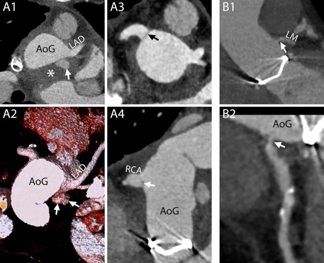
Figure 14.
Complications at the site of coronary arteries anastomosis and ostia. A1-A4, Different complications involving the two coronary arteries in the same patient. On a CT scan performed 1 month after a Bentall procedure, this 27-year-old male showed signs of complications at both coronary arteries. At the level of the suture line of the left main, a small collection of extravasated contrast material (A1 and A2, arrows) surrounded by a hematoma (A1, asterisk) was identified. The right coronary artery, that had to be reattached a second time to the aortic graft due to bleeding from the first suture, presented a hypodense line at the ostium (A3 and A4, arrows) that could be attributed to a small intimal flap or to an inward fold of the graft. No invasive or surgical treatment was undertaken. A1, Axial mage. A2, VR. A3, MPR perpendicular to the centerline of the aorta. A4, MPR parallel to the centerline of the aorta. B1-B2, Stenosis of the coronary ostium. On a routine follow-up CT scan performed 5 months after the procedure, this 60-year-old male showed a stenosis of the left coronary artery (B1 and B2, arrows). B1, MPR parallel to the centerline of the aorta. B2, CPR. AoG, aortic graft; LAD, left anterior descending; LM, left main; RCA, right coronary artery.

