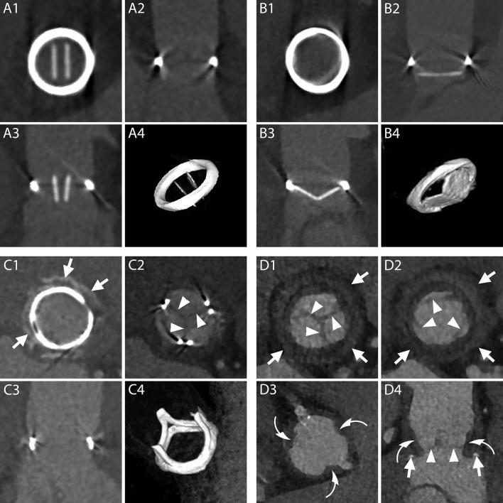
Figure 2.
Normal aspect of prosthetic aortic valves. A1–B4, CT aspect of the most commonly employed mechanical prosthetic valve (St. Jude, Abbott, Illinois, USA). The bi-leaflet valve is shown with open and closed leaflets respectively during systole (A1–A4) and diastole (B1–B4). A1, B1, MPR on planes perpendicular to the center line of the aorta at the level of the outer ring of the valve. A2, B2, MPR parallel to the centerline of the aorta and parallel to the direction of the leaflets. A3, B3, MPR parallel to the centerline of the aorta and perpendicular to the direction of the leaflets. A4, B4, VR of the valve. C1–C4, CT aspect of a stented biological valve during diastole. C1, C2, MPR on planes perpendicular to the centerline of the aorta respectively at the level of the most caudal (C1) and most cranial (C2) part of the metallic stent. In C1 the pledgets used to reinforce the sutures on the suture ring are visible (arrows). The biological leaflets are attached to the frame and when closed have an appearance similar to a native tricuspid aortic valve (C2, arrow heads). C3, MPR parallel to the centerline of the aorta. C4, VR of the valve. D1–D4, CT aspect of a stentless biological valve. D1, D2, MPR on planes perpendicular to the centerline of the aorta at the level of the suture ring of the valve (arrows) showing the three closed (D1) and open (D2) leaflets (arrow heads). D3, More cranially, at the level of the coronary arteries ostia, the outer frame of the valve appears as small hypodense irregularities (curved arrows) of the aortic contour protruding in the lumen. D4, An MPR parallel to the center line of the aorta shows the cusps of the leaflets (arrow heads), the outer frame of the valve (curved arrows) and the pledgets used to reinforce the sutures (arrows). MPR, multiplanar reconstructions; VR, volume rendering.

