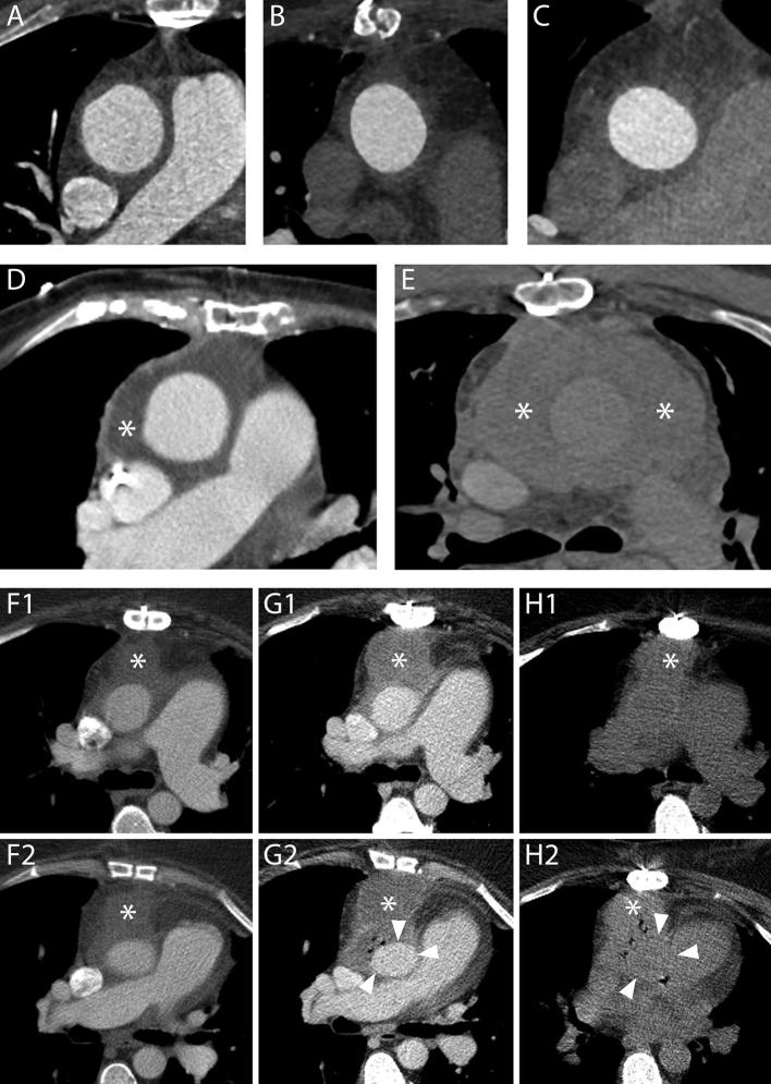
Figure 8.
Periaortic fluid in the form of stranding and fluid collections. A–C, Three different cases showing increasing amounts of periaortic fluid in the form of stranding. The scans were performed 55 (A), 69 (B) and 59 (C) days after an uncomplicated operation, respectively. In all cases, the patients had undergone an uneventful procedure and did not have any complications within the first year following the operation. D, A fluid collection with water-like content (asterisk) 12 days after a Bentall procedure in an 83-year-old female. The patient was reoperated 9 months later because of progressive dilation of the descending aorta. E, The day after a Bentall operation for type A dissection extending to the right carotid artery, this 61-year-old male presented a reduction of hemoglobin levels that prompted to investigate the source of bleeding. A CT scan was performed which highlighted the presence of a hematoma completely surrounding the aorta (asterisks). The patient died two days later for neurological complications. F1–H2, A 53-year-old female who had undergone a Bentall and a MAZE procedure four days before started complaining of posterior chest pain at the level of Th7. A CT scan (F1, F2) performed for the suspicion of spondilodiscitis or mediastinitis demonstrated a fluid collection (asterisks) with water-like density surrounding the aortic graft. Three days later, the patient had high fever and a new CT scan was performed to locate the focus of infection. Axial images (G1, G2) showed an increase in diameter of the previously reported fluid collection (asterisks) that had, at that time, clear characteristics of an abscess with air bubbles inside and slight compression of the prosthesis (arrow heads). Appropriate antibiotic therapy was started. The day after an unenhanced CT scan (H1, H2) demonstrated an increase in the air content of the collection (asterisks) albeit with an overall reduction in size and a restoration of the normal aortic graft appearance (arrow heads). The following day, the patient was reoperated and the collection drained.

