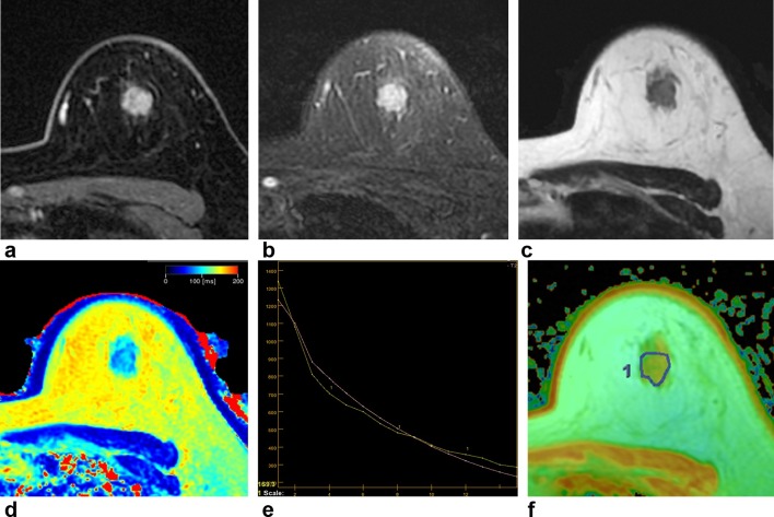
Figure 1.
A 47-year-old female with invasive ductal carcinoma in the left breast. Contrast-enhanced axial image (a) shows an enhancing mass with irregular shape and margin in her left breast 12 o’clock direction. T 2 weighted IDEAL fast spin echo image (b) shows an irregular mass showing high signal intensity in the same location. On synthetic T 2 weighted image (c) and synthetic T 2 map image (d), the T 2 relaxation time was 87 ms. On MESE T 2 mapping, T 2 relaxation time can be measured using 16 different TEs (e). The x-axis indicates TEs and y-axis indicates the signal intensity (e). On MESE T 2 mapping (f), the T 2 relaxation time was 77.9 ms showing difference of 9.1 ms compared to synthetic T 2 mapping. IDEAL, iterative decomposition of water and fat with echo asymmetry and least squares estimation; MESE, multi echo spin echo; TE, echo time.

